Written by Efrat Sasson PhD. Neuroimaging Scientist, Sagol Center for Hyperbaric Oxygen Therapy Center
The brain is a fascinating organ, and its complexity is gradually being revealed by scientists. Since longevity is a popular topic in modern society due to increasing life spans, aging of the brain is being carefully investigated by scientists.
The aging brain undergoes a vast amount of changes that influence everyday life and increase risk factors for many neurodegenerative diseases.
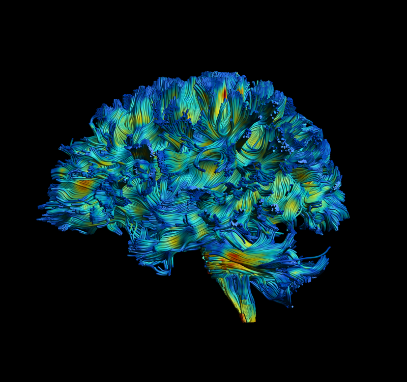
One of the brain tissues which draws a lot of attention in the medical community is white matter. The brain is essentially like a telephone network. Different functional regions are connected to one another by nerve fibers, allowing communication between them. Electrical signals pass through these fibers like telephone cables and the fibers are surrounded by a white fatty tissue called myelin that facilitates faster conduction.
During aging, there are different pathological changes in these wires, in the axons themselves and in the myelin surrounding them. These changes are even more severe in diseases and disorders such as Alzheimer’s.
But how can we visualize these changes?

Magnetic Resonance Imaging Sees White Matter
Most people know that getting an MRI means being slid into a big machine that provides high-resolution images of internal organs. But MRIs also can be used like a kaleidoscope. When twisting the kaleidoscope, you get different images and contrasts that are not only anatomical but quantitative. This is of great value to scientists.
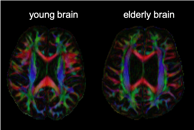
The technique that allows visualization of white matter is called diffusion MRI, or diffusion tensor imaging (DTI). The DTI technique illustrates white matter graphically and shows deterioration in white matter due to age. The regions of the brain responsible for memory and executive function (temporal and frontal regions) are those which deteriorate faster and more severely, according to a study published in the Neuropsychological Reviews.
Additionally, the white matter quantitative measures are correlated with cognitive performance. In a study published in Frontiers in Neuroscience, subjects who performed better in a mathematical task, had higher density of fiber tracts in a brain region known to be involved in mathematical calculation.
Amazingly, using this method has shown that the brain can change. In a study published in Nature Neuroscience: subjects were trained to juggle. The subjects were scanned before and after the training using DTI, and changes in the white matter were found in a brain region related to the complex task of juggling. In a study performed at Tel Aviv University, changes in the brain were even shown in the same day after memorizing a spatial track in a video game.
These studies demonstrate that the brain is not rigid. It is flexible and has the ability to change. These phenomena are called “brain plasticity.”
Changes in the brain are not a predestination. You can improve the brain even in aging, and imaging enables us to see even the little differences it makes.
